


A low retention micro-centrifuge tube improves sample recovery when a meaningful fraction of the sample remains on the wall after transfer, binds to the tube surface during contact, or experiences both losses. The strongest reasons to evaluate one are small working volumes, dilute analytes, repeated transfers, and storage before a sensitive assay. Selection should follow the observed loss mechanism: a tube that drains cleanly does not automatically preserve dissolved proteins or nucleic acids.
Before changing tubes, establish whether the problem is missing liquid, reduced analyte concentration, or reduced biological activity. These outcomes require different measurements. A nearly empty tube can still retain adsorbed molecules, while a visible droplet may contain analyte at the original concentration. Better recovery is demonstrated by the amount of usable analyte reaching the next step under the intended workflow.
Separate residual liquid from molecular adsorption
Liquid retention occurs when droplets or films remain on the inner wall, cap, or bottom after aspiration or pouring. Surface wetting, tube geometry, liquid viscosity, and pipetting technique all affect it. For a small aliquot, the liquid left behind represents a larger fraction of the starting volume. A surface designed to reduce wetting can improve drainage, although its behavior must be confirmed with the actual buffer.
Molecular adsorption removes analyte from solution through interactions with the tube surface. Proteins differ in charge, hydrophobic regions, and structural stability; nucleic acids differ in length, structure, and buffer environment. Polypropylene is widely used for microcentrifuge tubes, but the polymer name alone does not establish low-binding performance. Surface treatment, formulation, and manufacturing conditions also influence the interaction.
The distinction becomes useful when interpreting results. If the recovered volume falls while analyte concentration stays similar, residual liquid is a plausible explanation. If the recovered volume is comparable but concentration decreases, adsorption, degradation, or precipitation deserves investigation. If concentration remains stable while enzyme activity falls, a functional change has occurred that a volume measurement cannot reveal.
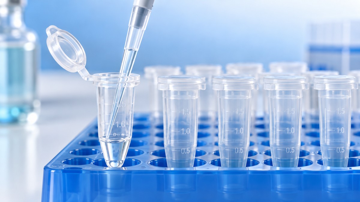
Conditions that make the tube surface matter
Low concentration increases the relevance of adsorption because a limited amount of surface-bound material can represent a substantial share of the available analyte. For proteins, abundant material in the sample can occupy binding sites and reduce the relative loss of a dilute target. A purified protein in a simple buffer therefore does not necessarily behave like the same protein in a complex biological matrix.
Small working volumes increase the wetted surface area relative to liquid volume for many tube configurations. An oversized tube can also spread sample across a larger wall area during mixing. Selecting an appropriate tube capacity and bottom geometry may improve handling before any surface modification is considered. Nominal capacity alone gives little information about how a small aliquot behaves near the tip of the tube.
Repeated transfers create fresh opportunities for loss. A workflow involving dilution, aliquoting, incubation, and movement into an assay vessel should be evaluated across that sequence. Testing only the first transfer can miss cumulative losses or attribute a downstream problem to the wrong container. Low-retention pipette tips also deserve controlled treatment because changing tips and tubes simultaneously obscures which component produced the improvement.
Longer contact times can expose adsorption that is absent during immediate transfer. Storage temperature, freeze–thaw cycles, and mixing affect sample stability as well as surface contact. Compare candidates at the actual holding time and temperature. A tube that performs well during rapid dispensing has not thereby demonstrated suitability for prolonged storage.
Choose the surface claim that matches the sample
“Low retention,” “low binding,” and “low adsorption” are not sufficient descriptions of a shared performance level. Product information should identify what was evaluated: residual liquid, protein binding, nucleic acid recovery, or another endpoint. Evidence obtained with water does not establish protein recovery, and evidence from one purified protein does not establish behavior for every enzyme, antibody, or peptide.
For protein work, examine whether the supporting test uses a relevant concentration, buffer, and contact period. Surface charge and hydrophobic interactions change with solution conditions. A treatment that improves recovery in a simple aqueous preparation can give a different result in a formulation containing salts, detergents, or stabilizers.
For DNA and RNA, distinguish adsorption from nuclease-related degradation. A low-binding claim does not establish freedom from DNases or RNases; these are separate product attributes. Fragment size and concentration matter when assessing recovery, and an assay optimized for one nucleic acid population may overlook selective loss of another.
Where documentation identifies a coating or surface treatment, establish compatibility with the intended solvents, temperature range, and downstream assay. Potential interference should be investigated through suitable blanks rather than assumed from the treatment type. Conversely, “uncoated” does not by itself prove better analytical cleanliness or recovery.
Mechanical suitability remains a separate requirement. The tube must tolerate the intended relative centrifugal force, rotor configuration, temperature, and chemical exposure. Use the specified force limit and its stated conditions; rotational speed alone does not describe the load without rotor radius. Cap closure also matters because evaporation or leakage can change apparent concentration and imitate a recovery problem.
Buffer composition can change the result
A surface that sheds water effectively may behave differently with a detergent-containing buffer. Surfactants alter wetting and interact with both analytes and surfaces. They can reduce some adsorption losses while changing drainage or introducing assay interference. Any benefit must therefore be judged in the final formulation, including additives introduced during dilution.
Protein carriers can reduce loss of a dilute target by occupying available binding sites, but they also change the analytical sample. They may be unsuitable for mass spectrometry, total-protein measurement, or applications requiring a defined composition. Adding a carrier solely to improve recovery should be assessed against the requirements of the next step.
Changes in pH and ionic strength affect molecular charge, solubility, and interactions with the tube wall. A falling soluble concentration after buffer adjustment could reflect precipitation rather than adsorption. Inspect for precipitate and use a measurement strategy that can distinguish soluble analyte from material removed during centrifugation.
Viscous solutions present another complication: slow drainage and pipetting error can dominate recovery even when adsorption is limited. Matching aspiration speed, dispensing technique, tip type, and settling time is necessary for a fair tube comparison. A candidate should not receive credit for an improvement caused by different handling.
Build a comparison around the actual transfer
Start with the current standard tube and one or more candidates, using aliquots from the same well-mixed preparation. Include the lowest relevant concentration and working volume, along with a routine condition. Keep the comparison manageable enough that each condition has adequate independent replicates; a large collection of single measurements provides weak evidence of consistent performance.
The reference should represent the amount available before the test exposure. An assay of the starting preparation, combined with a control designed to minimize additional handling, helps establish that baseline. Randomize processing order where timing or instrument drift could bias the result, and keep tube identity concealed during measurement when practical.
- Dispense matched aliquots using the same calibrated pipette and tip type. Record the working volume, formulation, and sample concentration.
- Apply the intended mixing, incubation, centrifugation, or storage conditions. Include cap contact if inversion occurs in the real workflow.
- Transfer the sample using a defined aspiration position and timing. Avoid special rinsing or extra centrifugation unless those steps belong to the routine method.
- Measure recovered volume and analyte concentration separately, then assess function or downstream assay performance where relevant.
Recovered analyte amount is the recovered volume multiplied by its measured concentration. Comparing that amount with the starting amount distinguishes a concentration change from actual recovery. Evaporation, for example, can increase concentration while leaving the total analyte amount unchanged. A concentration-only comparison would misinterpret that result.
Gravimetric measurements can support volume assessment when the balance resolution is adequate and liquid density is accounted for. Small-volume measurements require control of evaporation, exterior moisture, and timing. Fluorescence, absorbance, or amplification assays need a suitable working range and matrix controls so that buffer effects do not appear as tube-dependent losses.
Keep upstream sample preparation identical as well. If material is collected from Petri dishes, equipment such as the Manual Petri Dish Revolving Table, with its manual, dual-sided non-slip platform for 60–150 mm dishes, belongs to that earlier handling step. Dish handling should remain constant while tubes are compared, since unequal starting aliquots undermine the recovery calculation.
Interpret the result beyond a higher average
A useful improvement should exceed the variability of the measurement and appear under conditions that matter to the workflow. Report replicate spread alongside mean recovery. A higher average accompanied by erratic results may offer less practical value than a consistent result, particularly when samples approach an assay’s detection or quantification limit.
Acceptance criteria should be set before examining the comparison. These can address recovered analyte amount, variability, and an application-specific endpoint such as enzyme activity or amplification behavior. Avoid adopting an arbitrary universal recovery threshold: the acceptable loss for a concentrated stock differs from that for a scarce specimen used near the analytical limit.
When amplification is the endpoint, a shift in quantification cycle alone does not identify the mechanism. Extraction efficiency, inhibition, nucleic acid integrity, and amplification variability also influence the signal. Matched controls and an independent quantity measurement, where feasible, make the interpretation stronger.
If a candidate succeeds only with one buffer or concentration, define its use within that tested range. Extending the same conclusion to a different sample requires evidence. Confirmation with another manufacturing lot can also reveal whether the observed performance is reproducible beyond the initial evaluation.
Changing tubes is justified when the measured improvement survives the complete workflow and preserves the required analytical function. If recovery remains poor across tube types, the next investigation should follow the evidence toward pipetting, precipitation, degradation, evaporation, or downstream inhibition. Retain the conditions that produced the result with the tube selection, so later changes in formulation, volume, or storage prompt a relevant reassessment.
Related Posts
Online Message
PROFESSIONAL CONSULTATION
If you are interested in our products and want to know more details, please leave a message here, we will reply you as soon as we can.