


Can a hemacytometer deliver reliable bacterial counts, or do its limitations outweigh its convenience? Hemacytometers for microbiology can support direct counting under suitable conditions, but bacterial size, sample concentration, cell clumping, and microscope resolution all influence accuracy. Conventional chamber counts also do not distinguish viable from nonviable cells without additional techniques.
For technical evaluators selecting laboratory consumables, the question is therefore not simply whether bacteria fit inside the chamber. It is whether the chamber, microscope, sample preparation, and counting procedure together produce a result suitable for the intended decision. A quick estimate of a dense suspension demands different evidence from a viable-count result used for product release.
What a chamber count actually measures
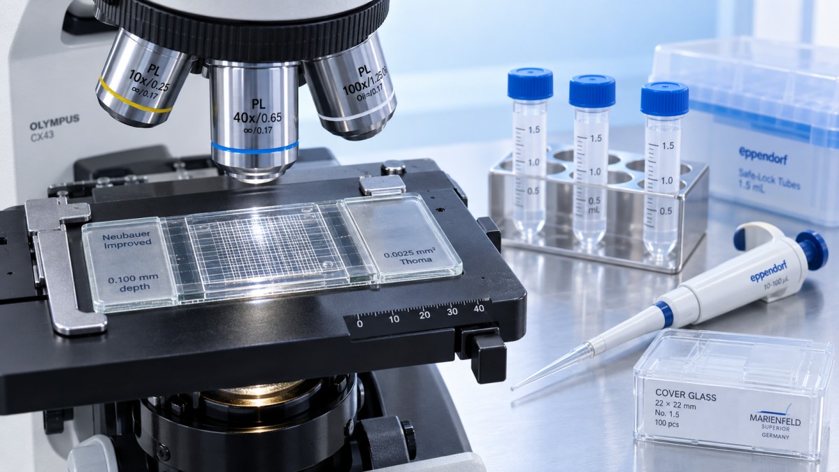
A hemacytometer, also spelled hemocytometer, defines a known volume above a ruled counting area. The operator counts visible objects within selected grid regions, then converts that count into a concentration using the chamber volume and any dilution factor.
For bacteria, the result is usually a direct microscopic count expressed as cells per milliliter. That description is valid only when individual cells can be identified and the counting rules specify how to handle chains, aggregates, and ambiguous objects. If a cluster cannot be resolved, calling it one “cell” creates a misleading result.
This measurement differs fundamentally from colony-forming units per milliliter, or CFU/mL. A colony may originate from one culturable cell or from several attached cells. Conversely, an intact bacterium visible under a microscope may be dead or unable to grow under the chosen culture conditions. Direct counts and plate counts can therefore disagree without either procedure necessarily being performed incorrectly.
Before purchasing a chamber, define the measurand: total visible cells, fluorescently labeled cells, culturable organisms, or another specified population. Equipment selection should follow that definition, not precede it.
Why bacteria challenge conventional hemacytometers
Small cells demand more than increased magnification
Many bacteria have dimensions on the order of a micrometer, with some considerably smaller. In an unstained wet preparation, their low contrast can make them difficult to distinguish from fine particles. Phase-contrast microscopy often improves visibility, but success still depends on objective resolution, illumination, chamber optics, and the sample matrix.
Higher magnification alone does not solve the problem. A high-power objective may have insufficient working distance for a particular chamber assembly, and the chamber’s optical surfaces may not suit the objective’s design. Do not assume that an oil-immersion objective compatible with ordinary slides is automatically compatible with a counting chamber. Confirm the manufacturer’s recommended optical configuration.
Chamber depth affects visibility and counting volume
A conventional improved Neubauer chamber commonly has a nominal depth of 0.1 mm. That depth is useful for many larger cells, but bacteria can occupy different focal planes through the liquid layer. An operator who counts only the easiest plane to see may miss a substantial portion of the population.
Bacteria-oriented chambers, such as certain Petroff-Hausser designs, commonly use a shallower depth, often 0.02 mm. A shallower layer can make focusing and enumeration more manageable, although it also reduces the volume represented by a given area. Always verify the actual chamber specification rather than transferring a calculation from another model.
Motility adds another complication. Moving bacteria may cross counting boundaries during observation, increasing the risk of duplicate counts or omissions. Any immobilization or fixation procedure must be evaluated for its effects on morphology, aggregation, and the intended measurement.
Concentration creates a narrow practical window
At low concentrations, too few cells appear in the sampled volume to support a precise estimate. At high concentrations, overlapping cells and crowded fields make individual identification unreliable. Dilution can resolve overcrowding, but it cannot rescue a sparse sample; concentration may be necessary and introduces its own recovery and bias concerns.
There is no universal bacterial concentration range suitable for every hemacytometer method. The useful range depends on chamber volume, cells counted, organism size, optical setup, and the laboratory’s acceptance criteria.
From grid squares to cells per milliliter
The calculation should be traceable to the chamber geometry:
Concentration = total cells counted ÷ total volume counted × dilution factor.
When area is expressed in mm² and depth in mm, their product gives volume in mm³. Since 1 mm³ equals 0.001 mL, unit conversion must be included. This is a common source of avoidable errors when laboratories change chamber types.
For example, consider a chamber with a verified depth of 0.1 mm. Counting a combined area of 1 mm² represents 0.1 mm³, or 0.0001 mL. If that area contains 200 individually resolved bacteria in a tenfold dilution, the estimated original concentration is:
200 ÷ 0.0001 × 10 = 2.0 × 10⁷ cells/mL.
This example demonstrates the arithmetic, not a recommended bacterial counting configuration. Changing the depth to 0.02 mm changes the volume fivefold, so the same count cannot use the same conversion factor.
Boundary conventions also matter. A common rule includes cells touching two designated borders and excludes cells touching the opposite borders. The laboratory must apply one consistent convention, define treatment of chains and clumps, and document the exact grid regions counted.
Counting more cells reduces random sampling uncertainty, but does not eliminate systematic errors. Under a simplified independent-counting model, relative counting uncertainty scales approximately as 1/√N. Counting 100 cells therefore corresponds to roughly 10% relative standard counting uncertainty; counting 400 reduces it to roughly 5%. Loading defects, clustering, and operator misclassification can make actual uncertainty larger.
When hemacytometers for microbiology are a reasonable choice
Direct chamber counting is most defensible when the suspension is sufficiently concentrated, the bacteria are readily distinguishable, and the objective is a total-cell estimate rather than a culture-based viability result. It can be useful for teaching, exploratory work, and monitoring relatively clean laboratory suspensions after suitable method verification.
The method becomes less attractive when the sample contains abundant debris, very small cells, extensive aggregation, or low bacterial concentrations. Complex clinical specimens can contain host cells and particles that interfere with identification. A chamber count alone generally cannot establish organism identity or support a diagnostic interpretation.
Optical density can be useful for tracking culture growth, but it is not a direct cell count. Its relationship to concentration changes with organism, size, aggregation, and instrument conditions. Use a relevant calibration rather than treating absorbance as a universal conversion to cells/mL.
What technical evaluators should verify before approval
A chamber specification is only one part of the evidence. For procurement and method qualification, evaluate the complete measurement system under representative laboratory conditions.
- Geometry and documentation: Confirm chamber depth, grid area, dimensional tolerances, and any supplied inspection or calibration documentation.
- Coverslip requirements: Use the specified chamber coverslip. Substituting an ordinary coverslip can alter the intended spacing or compromise seating.
- Optical compatibility: Demonstrate that the actual microscope and objective resolve the target bacteria across the counted area.
- Loading performance: Check for bubbles, overflow, incomplete filling, uneven distribution, and leakage into surrounding channels.
- Repeatability: Compare independent loadings and replicate counts, rather than repeatedly recounting one favorable field.
- Operator agreement: Test whether different trained analysts classify cells, debris, and aggregates consistently.
- Maintenance suitability: Verify cleaning and disinfection compatibility, resistance to routine handling, and replacement availability.
For an internationally sourced consumable, clear instructions and consistent specifications across shipments deserve particular attention. Distributor support is valuable when it provides traceable product identification, technical clarification, and reliable access to compatible replacement components—not simply an assurance that the chamber is “laboratory grade.”
A practical verification study, not a one-off demonstration
Start with representative organisms and matrices spanning the intended operating range. Include samples near the lower counting limit and samples requiring dilution. Agree on the counting area, border rules, mixing procedure, loading technique, observation timing, and rejection criteria before collecting comparative data.
Assess within-run repeatability, variation between operators, and between-day performance. Compare results with a reference procedure that measures the same biological quantity wherever possible. Agreement with another total-cell method addresses a different question from agreement with CFU/mL.
Dilution studies can reveal crowding or matrix interference. After correcting for dilution, concentrations should remain acceptably consistent within predefined limits. Poor agreement may indicate inadequate mixing, dilution error, adsorption losses, aggregates, or difficulty recognizing individual cells.
Set acceptance criteria according to intended use. A rough estimate for an exploratory experiment need not meet the same performance requirements as a quantitative result driving a regulated decision. Where a pharmacopoeial, regulatory, or contractual method applies, do not substitute chamber counting without establishing that the alternative is permitted and appropriately validated.
Bench organization supports consistency, but cannot fix optical limits
Small handling differences can undermine an otherwise sound procedure. Standardizing where clean supplies are stored, separating them from used chambers, and maintaining an approved cleaning sequence makes reproducible work easier.
For organizing compatible wash bottles in a designated clean area, the stainless-steel Wash Bottle Bracket, model ML4050, accommodates a maximum bottle diameter of 116 mm and measures 290 × 185 × 235 mm. Its handle supports movement, and its stated resistance to general chemicals is relevant to routine bench use. Compatibility with specific disinfectants still requires confirmation. This accessory supports organization; it does not improve bacterial detection or validate the counting method.
Cleaning agents, contaminated liquids, and reusable chambers must remain subject to the laboratory’s biosafety procedures. Never infer that an accessory or chamber is suitable for a particular decontamination process solely from its general material description.
The decision: suitable for a defined task, not a universal replacement
Hemacytometers can count bacteria when cells are visible, the sampled volume yields enough observations, and preparation and counting rules are controlled. Their convenience is real, but so are their limitations: small-cell resolution, focal depth, aggregation, manual variability, and the inability of conventional counts to establish viability.
For technical evaluators, the strongest purchasing decision connects chamber geometry and optical compatibility to a verified application. If the requirement is a defensible total-cell estimate in a suitable suspension, chamber counting may be practical. If the requirement is low-level detection, culturable counts, organism identification, or high-throughput precision, another method will often be the better fit.
Related Posts
Online Message
PROFESSIONAL CONSULTATION
If you are interested in our products and want to know more details, please leave a message here, we will reply you as soon as we can.