


To calculate cell concentration from a hemacytometer count, divide the number of cells counted by the chamber volume examined, then multiply by the sample’s dilution factor. For an improved Neubauer chamber with a depth of 0.1 mm, each 1 mm × 1 mm large square contains 0.0001 mL. When counting these squares, the calculation is cells/mL = average cells per large square × dilution factor × 104.
The 104 factor is not universal: it applies only to that counting area and chamber depth. Using smaller subdivisions, a different chamber design, or an incorrect dilution factor changes the result. A correct calculation also depends on a representative cell suspension, proper chamber loading, and consistent counting rules.
Match the calculation to the volume you actually counted
A hemacytometer measures concentration because its grid defines an area and its correctly positioned coverslip defines a depth. Together, these establish a known volume:
Counted volume = counted area × chamber depth
For one large square on a standard improved Neubauer grid:
- Area: 1 mm × 1 mm = 1 mm2
- Depth: 0.1 mm
- Volume: 1 mm2 × 0.1 mm = 0.1 mm3
- Conversion: 0.1 mm3 = 0.0001 mL
Dividing by 0.0001 mL is equivalent to multiplying by 10,000. This is the source of the familiar 104 conversion factor—not a correction for dilution or microscope magnification.
The general equation works with any verified chamber geometry:
Original sample concentration (cells/mL) = total cells counted ÷ total volume counted (mL) × dilution factor
If the counted area is expressed in mm2 and depth in mm, the equivalent equation is:
Cells/mL = total cells counted × 1,000 ÷ [total counted area (mm2) × depth (mm)] × dilution factor
Check the chamber’s instructions before choosing a conversion factor. Disposable counting chambers and alternative grid designs should not automatically be treated as improved Neubauer chambers.
Prepare a count that represents the suspension
Mix the suspension immediately before taking an aliquot. Cells can settle between sample preparation and chamber loading, so an aliquot collected from the top or bottom of an unmixed tube may not represent the whole suspension. Use a mixing method appropriate to the cells; vigorous handling that damages cells creates a different measurement problem.
If dilution is necessary, use accurate pipetting and record the volumes before counting. Mix the diluted sample again before loading. For viability measurements, follow the established staining protocol, including its timing requirements, because prolonged exposure to a dye can alter the apparent viable fraction.
For reusable chambers, fit the specified hemacytometer coverslip correctly before loading. An ordinary microscope coverslip may not establish the intended chamber depth. Introduce the sample at the loading edge and allow capillary action to fill the chamber without bubbles or overflow. Follow the manufacturer’s loading procedure for disposable designs.
Allow cells to settle into the counting plane according to the applicable procedure. Before recording numbers, inspect several grid regions. Obvious gradients, bubbles, extensive clumping, or cells moving through the field are reasons to correct the preparation rather than proceed with arithmetic.
Use the same square selection and boundary rule throughout
For many cultured-cell suspensions, an established protocol uses the four large corner squares of an improved Neubauer grid. Other methods use additional large squares or specified central subdivisions. The correct choice depends on cell type, density, and the laboratory procedure; what matters mathematically is knowing the total area examined.
Count systematically within each selected square, using a consistent scanning pattern to avoid missing or revisiting cells. Record each square separately rather than keeping only a running total. Individual counts make uneven distribution easier to detect.
Apply one boundary convention consistently. A common rule is to include cells touching the top and left boundaries and exclude cells touching the bottom and right boundaries. Use the grid lines designated by the chamber instructions, particularly where boundaries contain multiple lines. Do not switch inclusion rules between squares.
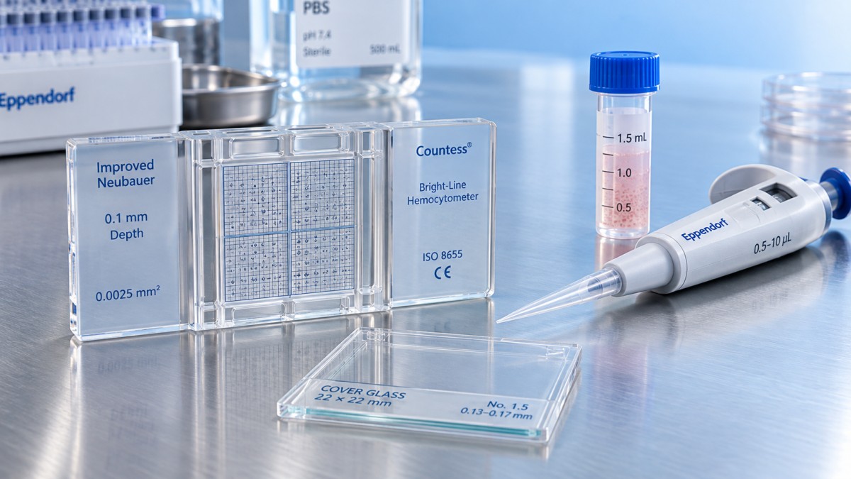
Define what qualifies as a cell before counting. Debris should not be included merely because it resembles a small cell at low magnification. If cells overlap so extensively that individual cells cannot be distinguished, additional dilution or an appropriate sample-preparation adjustment is needed. Treating an unresolved clump as one cell underestimates concentration; guessing its cell number is not a reliable substitute.
Worked example: converting four square counts into cells/mL
Assume a sample is mixed with an equal volume of diluent, giving a dilution factor of 2. Four 1 mm × 1 mm large squares are counted in a chamber with a depth of 0.1 mm.
The average count is:
180 ÷ 4 = 45 cells per large square
The concentration in the original suspension is:
45 × 2 × 104 = 900,000 cells/mL
The same result follows from the total-volume method. Four large squares contain 4 × 0.0001 mL = 0.0004 mL:
180 ÷ 0.0004 × 2 = 900,000 cells/mL
Use either approach, but do not combine them incorrectly. Multiplying the total of 180 cells by 104 without dividing by four would overstate concentration fourfold. The standard shortcut requires an average per large square, not the total across several squares.
Account for every dilution made before counting
The dilution factor is the final mixture volume divided by the volume of sample added:
Dilution factor = (sample volume + diluent volume) ÷ sample volume
Mixing 20 µL of sample with 20 µL of stain gives a factor of 2. Mixing 10 µL of sample with 90 µL of diluent gives a factor of 10. Undiluted material has a factor of 1.
Record actual volumes rather than relying on ambiguous ratio labels. “1:2” can be interpreted differently in informal notes, whereas “20 µL sample + 20 µL stain; dilution factor 2” is explicit.
For sequential dilutions, multiply the factors. If the original suspension is diluted tenfold and an aliquot is then mixed with an equal volume of stain, the overall factor is 10 × 2 = 20. Using only the staining factor would underestimate the original concentration tenfold.
Also identify which suspension the result describes. If cells were pelleted and resuspended in a new volume before counting, the measurement describes that resuspended material. Recovering the concentration of an earlier suspension requires additional volume and recovery information; a dilution correction alone does not account for cell losses during processing.
Separate viable concentration from total concentration
When using a suitable dye-exclusion method, record viable and nonviable cells separately within the same counted area. With trypan blue, viable cells are generally unstained and nonviable cells are stained, subject to the protocol’s classification and timing requirements.
Calculate each result from the corresponding count:
- Viable cells/mL = average viable cells per large square × dilution factor × 104
- Total cells/mL = average viable plus nonviable cells per large square × dilution factor × 104
- Viability (%) = viable cells counted ÷ total cells counted × 100
For example, suppose four large squares contain 160 viable cells and 20 nonviable cells after a twofold dilution. The average viable count is 40, giving 800,000 viable cells/mL. The total concentration is 900,000 cells/mL, and viability is 160 ÷ 180 × 100 = approximately 88.9%.
The dilution factor cancels in the viability percentage because both populations share the same dilution and counted volume. It does not cancel when calculating viable cells/mL. For procedures requiring a specified number of living cells, use viable concentration rather than total concentration. Dye exclusion indicates membrane integrity; it does not establish every aspect of cell function.
Recognize when the number is not reliable enough to use
A hemacytometer result can be mathematically correct yet unrepresentative. Large differences between squares or independently loaded chambers can reflect insufficient mixing, settling during sampling, uneven loading, clumps, or inconsistent classification. Compare equal counted areas and use the laboratory’s predefined acceptance criteria. If disagreement exceeds those criteria, investigate and reload rather than simply average away the discrepancy.
Counting very few cells also limits precision. Under an idealized random-counting model, relative counting uncertainty scales approximately as 1/√N, where N is the total number counted. A count of 100 cells therefore has roughly 10% relative counting uncertainty, while 400 cells gives roughly 5%. These estimates exclude pipetting, loading, and classification errors.
For sparse suspensions, count a larger verified area or reduce unnecessary dilution. For crowded suspensions, dilute further until individual cells can be distinguished, then update the dilution factor. Counting more cells improves sampling precision only when the sample and chamber loading are representative.
When combining counts from both sides of a chamber, sum the cells and the corresponding counted volumes. Eight large squares contain 0.0008 mL under the standard geometry. Alternatively, average all eight equal-volume square counts and use the 104 shortcut. If different areas were examined on each side, use total cells divided by total volume rather than a simple average of the two totals.
Use the concentration to prepare the required cell amount
Once the concentration is accepted, the sample volume needed for a target number of cells is:
Required volume (mL) = target cell number ÷ cell concentration (cells/mL)
At 800,000 viable cells/mL, obtaining 200,000 viable cells requires 0.25 mL, or 250 µL. Mix the suspension again before withdrawing that volume, since settling can make a later aliquot differ from the aliquot counted.
To dilute a suspension to a target concentration, use C1V1 = C2V2, provided the operation is dilution without cell loss. Preparing 2 mL at 200,000 viable cells/mL from a suspension containing 800,000 viable cells/mL requires 0.5 mL of suspension plus 1.5 mL of compatible medium. A target concentration above the starting concentration requires a concentration step, not dilution.
Retain the raw square counts, counted area, chamber depth, dilution volumes, and viable/nonviable classification with the result. Report the concentration with its units and avoid more significant figures than the measurement supports. Those records make a hemacytometer count traceable and help distinguish a calculation error from a sampling or preparation problem.
Related Posts
Online Message
PROFESSIONAL CONSULTATION
If you are interested in our products and want to know more details, please leave a message here, we will reply you as soon as we can.